 July
25
July
25
Tags
In a world of “blind imagination”: The science of Aphantasia
Picture yourself in your childhood home. Imagine your bedroom – your bed, perhaps some posters on the wall, and whatever other salient features you can conjure with your mind’s eye. Do you see yourself in it, or are you seeing it as though through your own eyes…or do you not “see” anything at all?
Aphantasia (coming from the Greek word phantasia meaning “imagination”) is the condition of being unable to evoke mental imagery – not merely of one’s memories, but of any sort of visual scene, real or imagined. Many congenital aphantasics (those born with the condition) spend much of their lives thinking that phrases like “picture yourself”, “visualize”, and “see with your mind’s eye” are meant figuratively rather than literally; often it is only in their teens or early adulthood when they learn that other people do, in fact, “see” things in their mind [1]. While aphantasia is in its very early days of public awareness and scientific research (the term “aphantasia” was only coined in 2015!), it has already sparked considerable interest and countless yet-to-be-answered questions.
The original Aphantasia
Though the name “aphantasia” may be new, the concept of poor (or even absent) mental imagery is not. The earliest descriptions can be traced to the late 1800’s by Sir Francis Galton (Charles Darwin’s cousin, worst known as a pioneer in eugenics). He expressed astonishment that a significant portion of his subjects, when given a set of instructions to imagine their bedroom or breakfast table, were flummoxed by the very nature of the question. One of his correspondents wrote to him, “These questions presuppose assent to some sort of a proposition regarding the ‘mind’s eye’ and the ‘images’ which it sees….. This points to some initial fallacy…… It is only by a figure of speech that I can describe my recollection of a scene as a ‘mental image’ which I can ‘see’ with my ‘mind’s eye’….. I do not see it…” [2]. Interestingly, Galton initially believed that this condition was particularly prevalent amongst scientists, going as far as to claim that “scientific men as a class have feeble powers of visual representation”. However, more recent data as well as reexamination of his own data refute this claim [3].
Moving into the twentieth century, differences in mental imagery abilities continued to be a source of great interest and a topic of much debate. Moreover, as cases of unique patients who suddenly lost their mental imagery came to light, further interest and debate was engendered over the neural and cognitive systems underlying visual mental imagery and how they differ from visual perception [4]. It wasn’t until the last decade, however, that public interest has really taken off, due in large part to one particular case study that used both psychological testing and neuroimaging technology to characterize one patient’s remarkable loss of “visual imagination”.
Aphantasia 2000: Patient MX
While most aphantasics have never possessed a “mind’s eye”, there have been rare cases of neurological patients who lost the ability to construct mental imagery. One such patient, “MX”, approached neurologist Dr. Adam Zeman and claimed that a few days after undergoing coronary angioplasty surgery, he was no longer able to visualize anything in his mind; his regular vision, however, was unperturbed. Even his dreams initially became entirely non-visual, although they eventually regained their visual content. ‘I can remember visual details, but I can’t see them,” he told the scientists. “I can’t explain that . . .. From time to time I do miss being able to see [in my mind]” [5].
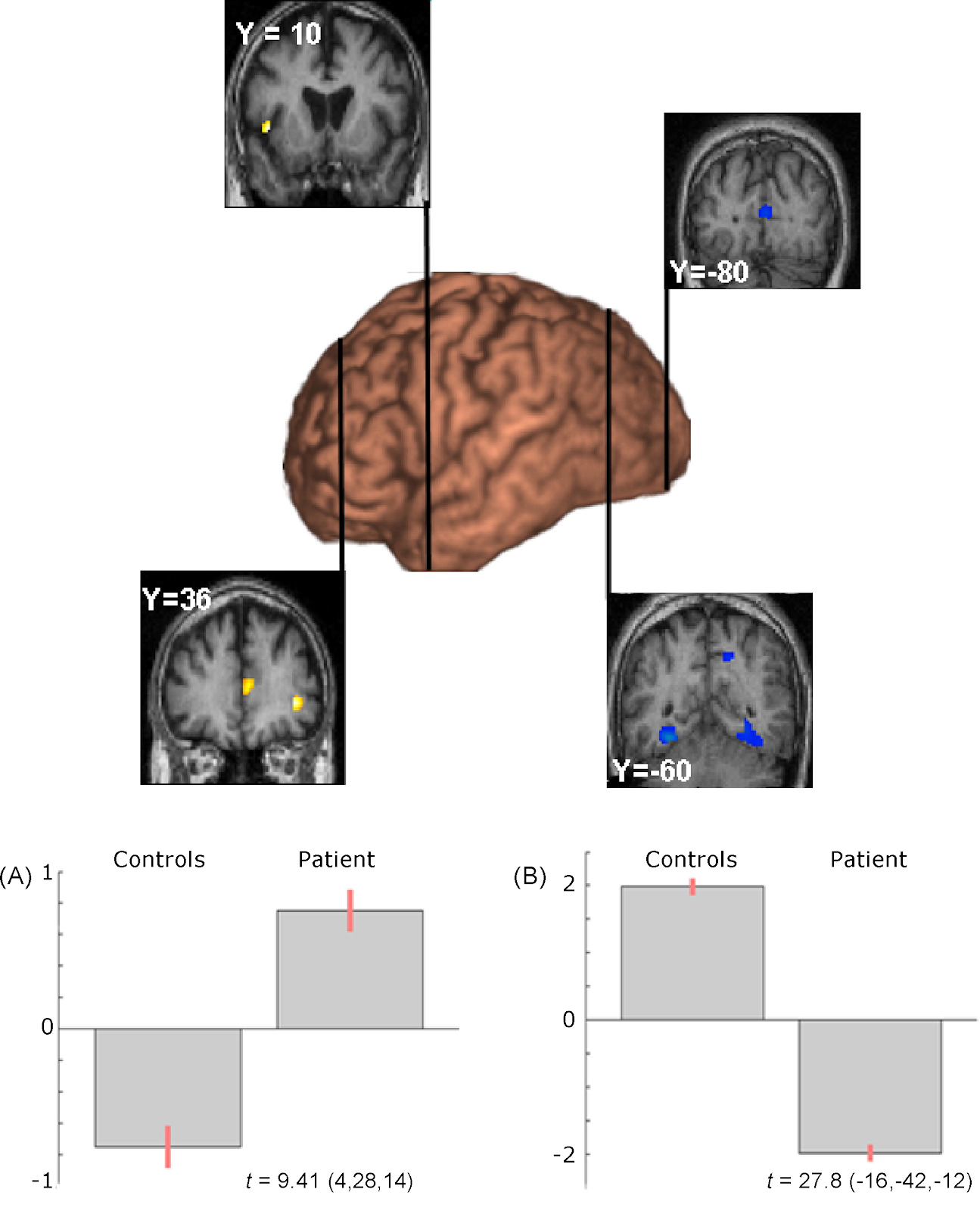
Curiously, while MX scored extremely low on self-report questionnaires of visual imagery, his scores on neuropsychological tests that supposedly assess visual imagery abilities were at normal levels. For instance, when asked to answer a number of questions about the shapes of different animals tails, most people would answer by visualizing the tails. MX, on the other hand, insisted that he wasn’t actually visualizing anything, yet he still aced the test. While MX excelled at this and many other visual imagery tests, some notable differences between him and similar but healthy control subjects emerged on a test of mental rotation. This test involves pictures of pairs of 3D objects comprised of multiple cubes. The subject has to respond whether the two objects are the same but rotated in 3D space relative to each other, or if they’re different objects. For most people, the amount of time it takes them to respond increases linearly with the angle of rotation. This would be expected if people were performing the task by visualizing the objects in their minds and mentally rotating them to see if they match up; a larger angle of rotation would thus take longer to make this comparison. MX performed somewhat slower on this task than the control subjects, but with higher accuracy. Interestingly, his response time was the same whether the objects were rotated by 40 or 140 degrees, suggesting that he was not using a mental rotation strategy. This was consistent with his own self-assessment, as he attested to solving the task by simply matching individual blocks and angles perceptually rather than attempting any sort of mental rotation.

While these results suggest that MX was performing these visuospatial tasks with some strategy other than mental imagery, Dr. Zemen and his team wanted to know if these processing differences were reflected in his brain activity. Previous studies on typical mental imagery have implicated a network of heightened neural activity in parts of the prefrontal cortex as well as posterior cortical areas that are involved in vision, with the interpretation that the prefrontal cortex serves to “reactivate” visual representations during mental imagery [6]. Since MX claims that he had lost access to these visual representations, one might expect that his visual cortex would fail to be “reactivated”. Remarkably, this is precisely what the researchers found. While MX’s pattern of brain activity was virtually indistinguishable from that of control subjects during basic visual perception (e.g., viewing pictures of faces), it was very different during visual imagery (e.g., imagining faces of famous people). Compared to control subjects who showed increased activity in prefrontal and posterior cortical regions during the facial imagery task, MX showed heightened activity in certain frontal regions and diminished activity in the same posterior cortical regions that were activated during face perception.
So how was MX able to perform so proficiently on tasks that supposedly assess visual imagery despite having a “blind imagination”? While it is difficult to know for sure, the researchers assessing MX hypothesized that visual information may be stored and represented in the brain in such a way that doesn’t necessarily require the brain’s visual network to be engaged. You might think of it as the distinction between visualizing the blue painted walls in your childhood bedroom and just knowing that the walls were blue. While most of us might engage our visual imagination to solve harder tasks – such as determining how well that Ikea bed will fit into the bedroom of your new apartment – those lacking visual imagery, like MX or congenital aphasics, might adopt some other, non-visual strategy to solve the same problem.
Aphantasia among us
While MX is a fascinating case of someone who experienced mental imagery before losing it entirely, it is more common that individuals with aphantasia have never known what it means to “see” something in your mind. And since it’s estimated that about 2% of the population experience aphantasia [7], it’s not even terribly rare; most likely you know someone with aphantasia (or perhaps that someone is you, and you are only learning of it for the first time!). Craig Venter, the famous geneticist known for first sequencing the human genome, has identified himself as having aphantasia. Beloved author and neurologist Oliver Sacks likely did as well, based on his own descriptions of “hardly see[ing] anything with my mind’s eye—at most, faint, evanescent images over which I had no control”, even though the name “aphantasia” was only just emerging by the time of his death. Interestingly, Sacks did note experiencing vivid, involuntary mental images while falling asleep and in his dreams, as well as drastically enhanced mental imagery for an approximately two-week period during the 1960’s following a hefty dose of amphetamines [8].
Even among those with congenital aphantasia, there seems to be a range of experiences. The theoretical physicist Nicholas Watkins wrote a fascinating article combining his own personal experiences of aphantasia and severely deficient autobiographical memory (SDAM), a review of some of the latest scientific research, and a hypothesis about how they relate to each other. Watkins suggests that SDAM, which is characterized by the inability to experience any sort of “mental time travel” or “Proustian”-like sensory memory, can be thought of as an extreme form of aphantasia. Though basic facts of one’s past are accessible, Watkins describes it as feeling like “the past really is another country to which I have no passport” [9]. On this hypothetical spectrum of aphantasias, “total aphantasia”, like that experienced by patient MX after his surgery, would be somewhat less extreme. This may be distinguished from SDAM in that these individuals can still experience the feeling of “mental time travel”, but without any visual component. Finally, individuals who might be characterized as having “voluntary aphantasia” cannot spontaneously conjure mental images on their own, but may still experience sudden, involuntary mental images, such as in the form of “flashes” and/or while dreaming [1]. Based on his own descriptions of his limited mental imagery, it seems likely that Oliver Sacks would have fallen into this latter category [9]. Though intriguing, the relationship between these forms of mental imagery deficiency is thus far no more than a hypothesis that has yet to be directly tested.
Among the general population, too, there is a wide range of visual imagination. For instance, on the opposite end of the spectrum from aphantasics are those with hyperphantasia who describe their mental images as nearly indistinguishable from real life. Then, of course, there’s everyone in between. Some of this heterogeneity can be captured by a widely used survey of visual imagery vividness (you can take it, too, as a participant for Dr. Zeman’s research!). Using this survey, Dr. Zeman and his research team divided a large sample of undergraduate research participants into “low-imagery” and “high-imagery” groups and conducted the same fMRI experiment as they did with patient MX. Intriguingly, the differences in brain activation during mental imagery between the low-imagery and high-imagery groups were very similar to those previously reported between patient MX and matched control subjects. The activation of multiple posterior cortical regions, including a particular area involved in face perception that was under-activated when MX attempted to imagine famous faces, were positively correlated with scores on the visual imagery questionnaire, whereas the activation of certain prefrontal regions were negatively correlated. Thus, these results are consistent with the idea that the engagement of some of these more posterior, visual areas are involved in the experience of mental imagery [10], although a causal relationship between these brain networks and mental imagery has yet to be conclusively established. Moreover, even the low-imagery participants in this study possessed some degree of visual imagination, i.e. were not aphantasic; differences in the patterns of brain activity between lifelong aphantasics and typical individuals has not yet been explored.
In the last 10 years – precipitated by Dr. Zemen’s published article about patient MX and the research into acquired and congenital aphantasia that followed suit – the concept of aphantasia and drastic inter-individual differences in mental imagery has exploded into the public consciousness. Since that article and the ensuing press coverage, Dr. Zemen’s group at the University of Exeter has reported receiving contacts from over 10,000 interested people, many falling into one of the extreme ends of the mental imagery spectrum [11]. As the public conversation about these inter-individual differences grows and resources for further scientific inquiry expand, we may begin to better understand humans’ internal experiences and their biological underpinnings.
References
2. Galton F. I.–statistics of mental imagery. Mind. 1880;os-V: 301–318. doi:10.1093/mind/os-V.19.301

Pingback: An Aphant's Look Into Aphantasia - J Living Full