Tags
Wait, do fish even have ears? Using zebrafish to study molecular mechanisms of hearing loss.
Hearing loss and vestibular dysfunction are among the most common disabilities in the world, affecting nearly one third of older adults. While much progress has been made researching the mechanisms underlying congenital deafness, the molecular and genetic mechanisms underlying adult-onset hearing loss and vestibular dysfunction are poorly understood. In the vast majority of cases of hearing loss vestibular disorders, loss of sensory hair cells in the inner ear is observed. The laboratory of Dr. Edwin Rubel at the University of Washington is focused on studying the development, plasticity, pathology, and potential repair of the inner ear and auditory pathways of the brain.
There are several ways that inner ear hair cells can be damaged, leading to adult-onset hearing loss: physical damage caused by excessive noise exposure, and pharmacological damage caused by environmental toxins, aminoglycoside antibiotics, and certain chemotherapy agents. Susceptibility to these potentially harmful factors is largely influenced by one’s genome. Due to the considerable amount of variation in the human genome among individuals, a single gene or set of genes is unlikely to be responsible for the broad spectrum of hearing loss disorders.
In order to uncover the many genes responsible for variation in sensitivity to ototoxic pharmaceutical drugs (those that cause damage to inner ear hair cells as a side effect), the Rubel lab collaborated with Dr. David Raible’s lab to develop a unique genetic and chemical screen using juvenile zebrafish mutants and known ototoxic agents. Using zebrafish as a model is advantageous because the hair cells on their lateral line are strikingly similar to mammalian and avian inner ear hair cells. In addition, the location of the lateral line hair cells on the outside of the body make it easy to perform pharmacological experiments as well as take time-lapse images of how the hair cells change. This method is described in detail in Owens et al (2008). When a zebrafish was exposed to the ototoxic aminoglycoside neomycin, a large percentage of their hair cells were lost.
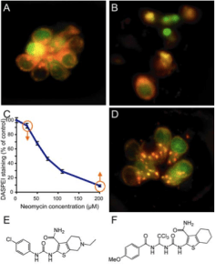
This figure demonstrates the screening method used for this study. Panel A shows a collection of zebrafish lateral line hair cells called a neuromast, which has not yet been treated by any drug. Panel B shows the result of exposure to 200µM neomycin: cell death of nearly all hair cells. The dose-response curve of neomycin is shown in C. When the cell is pre-incubated with PROTO-2, fewer hair cells are lost (D). The structure of PROTO-1 and -2 are in E and F.
By screening for both compounds and mutations that protected against this cell death, the researchers were able to investigate the underlying molecular mechanisms. A class of small molecules, benzothiophene carboxamides, was found to be protective against neomycin-induced cell death, specifically the molecules PROTO-1 and -2. In addition, several mutations were found to modulate sensitivity to neomycin, including one, sentinel, which substantially protected against cell death.
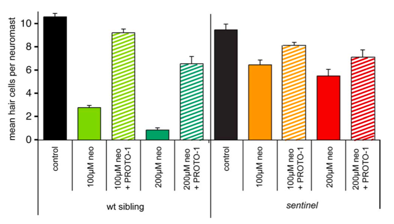
This figure summarizes the effects of PROTO-1 treatments and the sentinel mutation on neomycin-induced hair cell death. PROTO-1 and sentinel both partially protect against cell death. Importantly, PROTO-1 and the mutation protect more in combination than alone.
Intriguingly, PROTO-1 and -2 appear to act through a different pathway from the sentinel mutation; together, the mutation and the molecule were more protective against neomycin than either alone. Finally, the researchers found that PROTO-2 protected against neomycin-induced cell death in mouse utricle cultures, demonstrating that the insights gleaned from studying gene-drug interactions on zebrafish lateral line hair cells can be directly applicable to mammalian systems. In summary, Owens et al (2008) demonstrated a powerful method of screening for gene-drug interactions that will be incredibly important in studying how genomic variability affects drug efficacy.
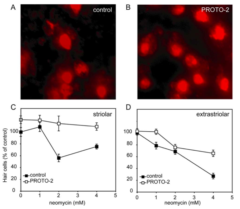
PROTO-2 protects against neomycin-induced hair cell death in cultured mouse striolar utricular hair cells.
To find out about the latest exciting discoveries in this line of research and others in the Rubel lab, please join us on Tuesday, January 8th at 4 pm in the CNCB auditorium for the UCSD Neurosciences Founder’s Lecture given by Dr. Ed Rubel, entitled “Fish in a dish: Discovering genetic and chemical modulators of inner ear hair cell death.”

Dr. Ed Rubel
Ethan McBride is a first year graduate student in the UCSD Neurosciences PhD program. Before coming to UCSD, Ethan worked in Ed Rubel’s lab for 3 years studying auditory neuroscience. This quarter he will be rotating in the lab of another PI named Ed, studying systems neuroscience with Ed Callaway at the Salk Institute.
Owens K.N., Santos F., Roberts B., Linbo T., Coffin A.B., Knisely A.J., Simon J.A., Rubel E.W. & Raible D.W. (2008). Identification of genetic and chemical modulators of zebrafish mechanosensory hair cell death., PLoS genetics, PMID: 18454195

I wish I had known about this talk earlier, rather than today, Jan 31, 2013. I might have come all the way up from Portland for it. I have a hearing loss and am especially interested in being in a clinical trial that could help me regenerate my hearing cells. Please let me know if there is one coming up. Thanks for posting this.