 August
01
August
01
Tags
Glued to Your Bed

As we know, sleep is essential for the proper functioning of our brain. Although sleep deprivation has never been found to be fatal in humans, a lack of sleep has been linked to neurodegenerative diseases such as Alzheimer’s, as well as delirium and hallucinations (1). This is why a lot of research delves into how the brain is affected by sleep and how it regulates our sleep (2;3). One recent study (7) found that the immune cells in our brain, known as microglia, may work with the brain to induce sleep by regulating neurotransmitters that help us stay alert and maintain attention. To understand how they do this, it is good for us first to have an understanding of the basic mechanisms of sleep and how sleep is impacted by different substances in the brain.
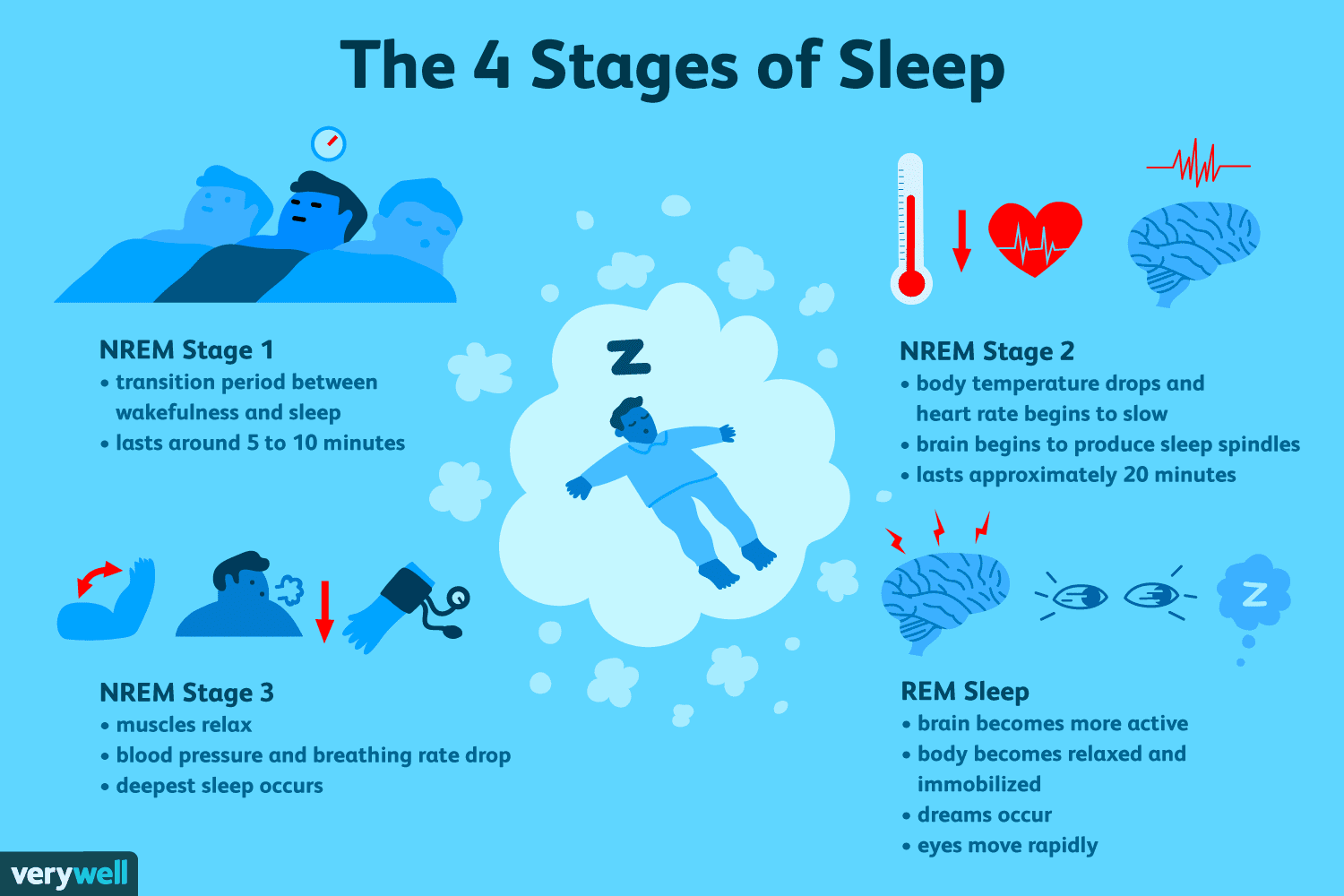
SLEEP PHASES
In the nervous system, sleep can be divided up into two categories, or phases: rapid eye movement (REM) sleep, and non-REM (NREM) sleep (4). REM and NREM sleep serve different purposes for the brain and body, and each has separate patterns of brain activity. During REM sleep, your brain activity is the same as and sometimes even greater than when you are awake; it is during this phase that your brain dreams, consolidates memories, and even regulates emotions. On the other hand, NREM sleep is when you enter the deepest sleep, with the slowest brain activity and a significantly decreased heart rate. It is during this phase of sleep that the body repairs and regrows tissues, builds bone and muscle, and strengthens the immune system. The NREM phase of sleep is also what researchers focused on to study microglia’s effects on sleep, especially the transition between wakefulness and NREM sleep.
NEUROTRANSMITTERS AND SLEEP
The brain communicates with itself and the body with special chemicals known as neurotransmitters (NT), and the regulation of one particular NT, known as norepinephrine (NE), is crucial for adequate sleep. In the brain, NE plays an essential role in the regulation of arousal (wakefulness), attention, cognitive function, and the stress response (“fight or flight”) (5). When it comes to sleep, the suppression of NE’s communication is what researchers saw when the research participants went from a wakeful state to sleep. Since NE plays a vital role in keeping us alert (wakeful) and directing our attention, the suppression of this chemical is important for us to get to sleep; intuitively, you can’t fall asleep if your mind is racing with ideas.

This isn’t the only part of the sleep equation, however. Believe it or not, the immune cells in the brain, known as microglia, actually help regulate (suppress) NE levels in the brain. Microglia play many roles in the brain: along with being the primary immune cells in the brain, they help clear the brain’s tissues of cellular debris and dead cells, secrete substances to help the other cells in the brain survive and flourish, and can even communicate with other cells to coordinate immune responses and regulate overall brain function. Because of their myriad functions, especially their ability to communicate with other cells via chemical messengers (e.g. NTs), microglia are also crucial for regulating sleep. Understanding the critical roles that NE and microglia play in the regulation of sleep is essential for understanding the recent research regarding the interaction of microglia and NE to influence sleep.
NEUROTRANSMISSION
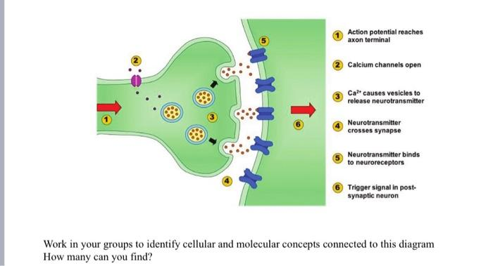
Neurons are the fundamental units of the brain, and they communicate with each other through a process known as neurotransmission. In neurotransmission, neurons release neurotransmitters into the space between neurons, known as the synaptic cleft, and these chemicals interact with receptors on the receiving neuron to produce a response in it. Neurotransmission is how the brain allows us to interact with the world and carry out actions.
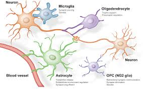
However, neurons aren’t the only cells in our nervous system that communicate using neurotransmission. The other class of cells in the brain, known as glia or glial cells, can also use neurotransmission to communicate with neurons. Glial cells are the support cells in the brain (“glia” is the greek word for “glue”), and there are many types of glial cells in the brain. Microglia is one type of glia in the brain, and recent research has found that microglia regulate neurotransmission to aid in sleep just as much as neurons do.
The basic process of neurotransmission is facilitated by the movement of chemicals known as ions into and out of a cell. One of the ions that is essential for the signal of one neuron to be released to another is calcium (Ca2+). We can measure the amounts of Ca2+ inside and outside of cells as one way to see how much the cells are communicating with each other, using a technique known as 2-photon calcium imaging. During 2-photon calcium imaging, researchers measure the calcium levels inside neurons by using fluorescent molecules that bind to the calcium, and an increase in the intensity of this fluorescence means that there is more calcium, thereby indicating increased neurotransmission (6). In the present study, researchers used this technique to measure the activity of microglia in mice. Elevated levels of Ca2+ within microglial cells indicated they were more active because Ca2+ is what begins the cascade of events of neurotransmission within a cell.
However, the elevated levels of Ca2+ that were seen in this study occurred at the same time as there was a significant reduction in NE release. How does that make any sense, you may ask? Well, this happened because microglial cells inhibited the activation of neurons (7). One of the effects of this is a reduction in neurotransmission, and thus a reduction in NT release, such as NE. Thus, researchers were able to link the inhibition of activity and reduction of NE release to better sleep by looking at the changes in the levels of Ca2+ within the cells. By looking at these changes during the transitional periods of wakefulness and NREM sleep, they could see if there were any meaningful differences in the concentrations of Ca2+. Most importantly, in both transitions, wake→NREM and NREM→wake, changes in the concentrations of intracellular microglia Ca2+ and extracellular NE showed opposite responses. When going from a wakeful state to NREM, intracellular Ca2+ increased, and extracellular NE decreased; conversely, NREM→wake saw a decrease in Ca2+ levels and an increase in NE. All of this shows us that the interaction between microglia and NE may be essential for adequate sleep.
Good Night
Recent research, such as the study discussed in this article, underscores the integral role of microglia, the brain’s immune cells, in modulating neurotransmitters crucial for sleep. By understanding neurotransmitter dynamics and microglial activity, we can begin to investigate how we can better help those who suffer from insomnia and other sleep disorders. In fact, some of the current treatments for insomnia already use some the mechanisms discussed in this article to aid sleep: one medication used to treat insomnia, known as Trazodone, is believed to aid in sleep because of properties that impede NE from taking effect. With research like this study, even more innovative therapeutic interventions aimed at combating sleep disorders are made possible.
References
- https://www.betterhealth.vic.gov.au/health/conditionsandtreatments/sleep-deprivation
- https://pubmed.ncbi.nlm.nih.gov/9322273/
- https://psycnet.apa.org/record/1965-13655-001?doi=1
- https://en.wikipedia.org/wiki/Sleep_cycle
- https://www.ncbi.nlm.nih.gov/books/NBK540977/#:~:text=First%20identified%20in%20the%201940s,cognitive%20function%2C%20and%20stress%20reactions.
- https://www.ncbi.nlm.nih.gov/pmc/articles/PMC10732251/#:~:text=In%20vivo%20two%2Dphoton%20calcium,molecules%20that%20bind%20to%20calcium.
- https://www.nature.com/articles/s41593-023-01548-5

You must be logged in to post a comment.