Tags
Primate Visual Space: The Entorhinal Frontier
Throughout the course of human history, great metaphorical emphasis has been placed on developing an understanding of our “place in the world.” Although this proverbial construct refers more to a sense of self-efficacy, it underscores the inarguable importance of determining our position as it relates to the environment around us in producing proper behavior. Research into the neural mechanisms underlying determination of place in relation to the individual has successfully delved into this field (no pun intended), elucidating an essential role for specific cell types in the hippocampal formation in processing location-relevant information.
Despite comprehensive research revealing the existence of place and grid cells expressly for this purpose in rodent models, the mechanisms of location-dependent learning and memory in the primate brain remain somewhat of a mystery. Thankfully, Dr. Elizabeth Buffalo and colleagues at Emory University have committed to tackling this very question.

Dr. Elizabeth Buffalo
In a recent paper, Buffalo and colleagues explored spatial representation in the monkey entorhinal cortex and hippocampus using a free-viewing visual memory task called the visual preferential looking task (VPLT). Monkeys were head fixed in a chair and shown novel images of fixed reference frame, twice each, on a computer monitor. A laminar electrode array was placed in the entorhinal cortex of each of the three monkeys in order to record spikes and local field potentials, allowing for recording from a total of 342 neurons in the hippocampal formation. Data regarding gaze location was determined using an infrared eye-tracking system (ISCAN).
The authors found that the monkeys explored the images, which consisted of various complex elements including abstract art, landscapes, animals, and people, with a series of dynamic fixations that coincided with increases in entorhinal cortical firing. Conversely, changes in hippocampal activity were significantly more variable.
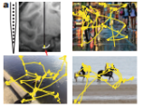
Fig 1. Examples of images shown and imposed representations of dynamic fixations
Buffalo and colleagues identified these cells as grid cells, and observed a significant presence in the posterior EC, but not in the hippocampus. Moreover, grid cells were located in both superficial and deep layers of EC, indicating a possible role in processing both input and output from the hippocampus. These results were consistent with expectations based on analogous structures and projections in the rodent brain (i.e. MEC). The authors additionally concluded that firing rate was independent of stimulus content.

Fig 2. (Middle) Grid cells and Border cells both qualitatively identified in primate EC. (Right, Left) Autocorrelations for representative cells of these types.
Buffalo and colleagues also noted a significant population of EC neurons that increased firing rate when gaze was located near the edges of stimuli. Qualitatively, these cells matched the border score criteria used in rodents to identify border cells, indicating that the visual map of space in the entorhinal cortex of primates may similarly be anchored in a framework in which the boundaries of stimuli serve as landmarks.

Fig 3. Reductions in firing upon exposure to repeated stimuli were more pronounced in anterior regions of EC (grey areas demarcate significant response reductions)
At more anterior locations, larger proportions of visually responsive neurons showed a reduction in firing rate in response to repeated stimuli, with the magnitude of this reduction increasing progressively, insinuating a role in recognition memory. This memory-linked response seemed to be independent of spatial representation, as while grid cells in anterior regions of EC were more likely to produce a memory response, the density of this cell type gradually declined along the anterior axis, with absolutely no grid cells located beyond the posterior 50% of the EC.

Fig 4. The probability of spiking for EC grid cells phase locked to theta oscillations
Finally, Buffalo and colleagues determined that the EC exhibited theta oscillations that resemble those found in primate hippocampus, and that the grid cells appeared phase-locked with the trough of the local field potential theta, suggesting theta modulation of these cells. However, firing rate maps of grid cells during theta and non-theta bouts did not appear significantly different, requiring adaptation of the current grid cell models basing their function on interactions of oscillations.
Buffalo’s work is both fascinating and imperative for attaining a fundamental understanding of primate learning and memory. In addition to detailing and localizing the qualities of the primate visual map, Buffalo confirmed that primates can form spatial representations during visual exploration at a distance. This discussion barely scratches the surface of the incredible work the Buffalo lab performs to elucidate the complexities of learning and memory in primates. Join us on Tuesday, March 5th at 4 pm in the CNCB auditorium for what is sure to be an informative and exciting lecture by Dr. Elizabeth Buffalo!
Landon Klein is a first year in the UCSD Neurosciences Graduate Program. He possesses a strong interest in drug mechanisms and addiction. Landon is currently rotating in the Alcoholism and Addiction Lab of Dr. Marisa Roberto at The Scripps Research Institute. Landon insisted on eating buffalo chicken last night in honor of Dr. Buffalo’s groundbreaking work.
Killian N.J., Jutras M.J. & Buffalo E.A. (2012). A map of visual space in the primate entorhinal cortex, Nature, DOI: 10.1038/nature11587

Wow, that’s what I was exploring for, what a stuff! present here at this blog, thanks admin of this website.