 June
12
June
12
Tags
Pattern separation gone awry: the dentate gyrus and schizophrenia
[Image Source: Sebastian Seung via http://connectomethebook.com/.]
Since the discovery of patient H.M. in the 1950s (see this post from October 2013), scientists have known that the hippocampus, a seahorse-shaped structure located in the medial temporal lobe, is crucial for the successful formation of new memories. The mammalian hippocampus is characterized by several distinct regions, each with a different function, including areas called the cornu ammonis fields (CA1, CA2, CA3) and the dentate gyrus (see Figure 1).
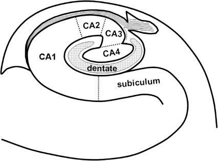
Figure 1. Schematic of the hippocampus. Source: Yang et al., 2008 [9].
Dentate gyrus: a pattern separator
The dentate gyrus, a structure named after its “toothy” appearance due to the presence of many small blood vessels in the region, is thought to be important for distinguishing between similar patterns or items in memory [2]. Pattern separation, broadly defined, can refer to distinguishing any two memories based on their representational (or neuroanatomical) “pattern”—or “…transforming similar input representations into highly dissimilar output representaions” [2, p. 293]. For example, where you parked your car last night might be really similar to where you parked it two nights ago, but the dentate gyrus might be able to help you distinguish between the two competing memories by separating the two patterns of activation associated with the two memories.
Other regions of the hippocampus (including CA1 and CA3) are thought to be important for pattern completion, or accessing a complete representation in memory from only partial input. These functions may complement the dentate gyrus’s ability to do pattern separation, for instance, selecting a memory representation from other competing representations.
The dentate gyrus is one of the few brain regions which continues to produce neurons (a process called neurogenesis) throughout adulthood (both in humans and in other mammals, such as rats). Recently, neuroscientists have hypothesized that neurogenesis may play a crucial role in differentiating between different events (or episodes) that animals experience [3]. Rangel and colleagues propose that neurogenesis can act as a time stamp, with newer neurons being associated with more recent events. Physical differences in dentate gyrus neurons at the time of encoding new information (as a function of their age) may help differentiate between events that are separated by longer periods of time. That is, events that occur closer together in time might be encoded by groups of cells which differ in their age.
To test this hypothesis, the researchers recorded neurons from dentate gyrus in rats while the rats were placed in similar environments with a long separation in time between exposures to each environment. They found that many place cells (neurons which fire relative to a specific place in an environment, regardless of the absolute coordinates; see Figure 2) in the dentate gyrus were sensitive to only one of the three contexts, suggesting that the dentate gyrus may discriminate between patterns based on timing, in addition to other types of cues. That is, the dentate gyrus could use time as a strong cue in separating between similar events.
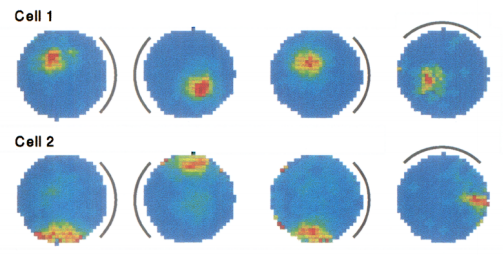
Figure 2. As the distal cue (represented by the arced line) changes position, the resulting firing pattern of neurons from hippocampus responds based on the location of the animal in relation to that cue. Cell 1 always preferentially responds (with greater response shown in warmer colors like red and yellow) when the animal is slightly to the left relative to the cue; cell 2, when the animal is to the right of the cue. Source: Knierem et al., 1995 [10].
Schizophrenia is often characterized by thought disorder, a symptom associated with abnormal patterns of activating meaningful information in (semantic) memory. Thought disorder is in turn often accompanied by incomprehensible speech, also known as “word salad,” where many inappropriate connections between context-inappropriate but related words may be present. For instance, one recent textbook [4] provides the following example:
“I don’t want to go to jail. Genesis, Revelations. April fools! Birds fly in. FoodMart birds. Take the bus to jail, take the birds to FoodMart.”
Relatedly, in some patients with schizophrenia, meanings of homophones that are irrelevant to the context are thought to be processed more strongly than in healthy individuals; that is, patients have difficulty inhibiting contextually irrelevant meanings of words [5].
The volume of the hippocampus is lower in schizophrenic patients bilaterally (that is, in both brain hemispheres) [6], and abnormal activation in the hippocampus during word memory tasks has also been observed in schizophrenic patients during functional MRI scans, particularly in the left hippocampus [7]. Finally, post-mortem studies of the hippocampus in schizophrenic patients has provided evidence that a marker for neurogenesis in the dentate gyrus is linked with schizophrenia [8]. That is, patients with schizophrenia may exhibit a reduced ability to generate new neurons in this brain region compared with healthy controls. Given that the dentate gyrus is thought to be important for successful pattern separation in humans [2], as well as in animals, this finding may indicate that dysfunction of the hippocampus, and the dentate gyrus, in particular, plays a role in some of the cognitive dysfunction in thought disorder.
One group of researchers [1] has suggested a model of hippocampal dysfunction in schizophrenia in which reduced dentate gyrus function changes the plasticity (that is, the ability of neurons to change their functional connectivity with one another) of the downstream hippocampal region CA3. The reduced functionality of the dentate-gyrus-to-CA3 pathway may reduce the ability of the dentate gyrus to function as a pattern separator while increasing the influence of CA3 as a pattern completer. If this is the case, then inappropriate associations and false memories or inappropriate representations may be activated, both at the initial instantiations of encountering an item and also at the point of accessing that item in memory. At an extreme end, this dysfunctional pathway could lead to psychosis.
Though still somewhat speculative, research in animals and humans has begun to suggest biological pathways that might lead to a better understanding of psychiatric disorders such as schizophrenia. In turn, this understanding of dysfunction in memory in psychiatric disorders can shed light on the basic neurobiology underpinning some of the most complex cognitive functions of human life.
References
[1] Tamminga, C.A., Stan, A.D., & Wagner, A.D. (2010). The hippocampal formation in schizophrenia. American Journal of Psychiatry, 167, 1178-1193.
[2] Azab, M., Stark, S.M., & Stark, C.E.L. (2014). Contributions of human hippocampal subfields to spatial and temporal pattern separation. Hippocampus, 24, 293-302.
[3] Rangel, L.M., Alexander, A.S., Aimone, J.B., Wiles, J., Gage, F.H. Chiba, A.A., & Quinn, L.K. (2014). Temporally selective contextual encoding in the dentate gyrus of the hippocampus. Nature Communications, 5.
[4] Compton, M.T. & Kotwicki, R.J. [Eds]. (2006). Responding to Individuals with Mental Illnesses. Sudbury, MA: Jones and Bartlett Publishers.
[5] Kiang, M., Kutas, M., Light, G.A., & Braff, D.L. (2007). Electrophysiological insights into conceptual disorganization in schizophrenia. Schizophrenia Research, 92, 225-236.
[6] Bogerts, B., Ashtari, M., Degreef, G., Alvir, J.M.J., Bilder, R.M., & Lieberman, J.A. (1990). Reduced temporal limbic structure volumes on magnetic resonance images in first episode schizophrenia. Psychiatric Research: Neuroimaging, 35, 1-13.
[7] Weiss, A.P., Schacter, D.L., Goff, D.C., Rauch, S.L., Alpter, N.M., Fischman, A.J., & Heckers, S. (2003). Impaired hippocampal recruitment during normal modulation of memory performance in schizophrenia. Biological Psychiatry, 53, 48-55.
[8] Reif, A., Fritzen, S., Finger, M., Strobel, A., Lauer, M., Schmitt, A., & Lesch, K.-P. (2006). Neural stem cell proliferation is decreased in schizophrenia, but not in depression. Molecular Psychiatry, 11, 514-522.
[9] Yang, Y., Kim, S., & Kim, J.H. (2008). Ischemic evidence of transient global amnesia: Location of the lesion in hippocampus. Journal of Clinical Neurology, 4(2), 59-66.
[10] Knierim, J.J., Kudrimoti, H.S., & McNaughton, B.L. (1995). Place cells, head directions cells, and the learning of landmark cues. The Journal of Neuroscience, 15(3), 1648-1659.

Pingback: How Does Exercise Improve the Brain? | NeuWrite San Diego
Pingback: The plastic brain | NeuWrite San Diego