 April
26
April
26
Tags
Lymph, glymph, sleep, & sickness
 Consider the word “lymph.” What comes to mind? To me, “lymph” sounds like a viscous liquid that might ooze out of the orifices of some terrifying wounded creature. Or perhaps your mind jumps to the term “lymph nodes”, conjuring images of little knobs in your neck bulging with infection, sometimes growing so large that they are detectable by a doctor’s touch. In reality, lymph is far less unpleasant than it sounds. In fact, the term lymph comes from Lympha, the name of a Roman goddess of fresh water. I would not necessarily say that lymph is fresh water, but it is primarily water, with a few small proteins and fats dissolved in it. It is constantly moving through us, even when we are in perfect health, and the proper functioning of our bodies depends on its cycling.
What is lymph and why do we need it?
 When we think of a fluid cycling through us, we of course first think of blood, not lymph. Blood is important for bringing oxygen and nutrients to all the cells of our bodies. But how does the actual exchange occur? Oxygenated blood from the heart flows through arteries, which branch into smaller capillaries, and oxygen can diffuse across the capillary walls. In most of the body (not the brain, but I will get back to that), some of the water and small molecules from the blood seep out of little holes in the walls of capillaries, bathing your body cells in “interstitial fluid” (ISF). ISF provides the cells with essential nutrients. Although this liquid comes from the blood, it is colorless because the red blood cells that give blood its color remain within the blood vessels, along with larger molecules that cannot fit through the gaps in the capillary walls. While some of the water flows back into the blood vessels by osmosis, a significant amount (~3 liters per day!) does not. Your bloodstream would dry up quickly if it continued losing so much liquid on a daily basis. The solution? The lymphatic system! The lymphatic system does a few things, but one of its most important jobs is getting all of this liquid back to the bloodstream.
 Special vessels called “lymphatic vessels” take up the ISF, and that’s when it technically becomes “lymph.” In addition to water and small molecules, lymph vessels will also take up any bacteria or virus that might be hanging out in your tissue. Lymphatic vessels throughout the body transport the fluid from your various organs to lymph “nodes”—basically little check-in points for lymph that are scattered throughout the body. If there is a suspicious stranger (like bacteria) in the lymph, white blood cells specifically made to attack that bacteria will multiply in the nodes and wage war, essentially filtering any infectious agents out of the lymph. This rapid multiplication of white blood cell warriors is the reason a doctor might feel for swollen lymph nodes as a sign of infection. Eventually, lymphatic vessels empty the filtered fluid into large veins near the heart, back into the blood circulation from which it originally escaped.

The brain, special as always
What does this have to do with neuroscience? As is so often the case, the brain is an exception to the general process explained above—there are no lymph vessels in the brain! Since there are no lymph vessels, technically there is no “lymph” either, but there is still blood flowing through blood vessels and ISF in which all of your brain cells float. This presents a problem. Neurons are incredibly metabolically active, thereby generating a lot of metabolic waste, and they are very sensitive to this waste since they require a very precise extracellular environment to properly communicate with each other. Given these facts, you’d think the brain would be chock full of lymphatic vessels, not totally without them!
Until quite recently [1], the details of fluid flow and waste management in the brain in the absence of specialized lymphatic vessels presented a conundrum. And while there is still plenty that we do not understand, in the last six years, neuroscientists have made exceptional progress in understanding the process of brain fluid exchange. The research is still new and somewhat controversial, so the details as described below may be refined as the research progresses. That said, what scientists have learned has far-reaching implications, including why we sleep and why aging brains are so vulnerable to disease.
To follow these discoveries, we need to dive a bit further into brain anatomy and the different types of fluid in and around the brain.
Fancy fluid flow

Rotating 3D view of the ventricles, where CSF is produced.
In addition to blood and ISF, there is cerebrospinal fluid (CSF), which is generated in the ventricles (large fluid-filled spaces in the brain). CSF has specific ionic concentrations, and it fills the ventricles and flows around the brain, surrounding it on the outside. Recently, scientists discovered that there is a constant fluid exchange between this special CSF produced by the brain and the ISF bathing brain cells. It is the force of this flow that is now thought to power waste clearance from the brain. But how?
Interestingly, starting from the surface of the brain, CSF flows into the brain along the outsides of arteries and capillaries, propelled by contractions of the same smooth muscle cells that facilitate blood flow. The CSF does not mix and mingle with the blood as it flows along the vessel walls because of the blood-brain barrier (capillary walls in the brain are far less permeable than in the periphery!). On the other side of this CSF tract is a wall of astrocyte endfeet. Astrocytes are star-shaped glial cells that reside only in the brain and are absolutely essential for the development and maintenance of neuronal connections and proper brain function. Their cell bodies lie amongst neurons, but some of them send out long thin processes, the ends of which (“endfeet!”) form a sheath around the brain’s blood vessels. As CSF flows between the capillary walls and the astrocyte endfeet, it can enter the brain tissue through special channels in the astrocytes (the “AQP4” channels seen in the diagram). When the CSF (water with a particular combination of ions) flows in and around the cells, mixing with cell nutrients and waste, it is then referred to as ISF.

CSF follows the arrows. It flows along the outsides of arteries and capillaries on its way into the brain. It can flow from the space next to the blood vessels through AQP4 channels in astrocytes, and out into the ISF bathing neurons. This flow pushes some metabolic waste products (“interstitial solutes” from the old ISF into a similar channel next to the vein. This flow eventually empties into the peripheral lymphatic system. Public domain image: https://en.wikipedia.org/wiki/File:Glymphatic_system_schematic.jpg
The pressure of CSF entering the space around neurons pushes some of the ISF into a similar space along large veins carrying blood back to the heart. Veins don’t pulse with the same strong pressure that arteries do, so the CSF-ISF flow is unidirectional, thereby washing out many of the brain’s waste products. Essentially, fresh CSF pushes out old ISF. (And by “old” I mean a few hours–the entire volume of CSF is renewed about every 6 hours in humans!). From this space next to the veins, the old ISF eventually empties into the peripheral lymphatic system. Because this fluid flux acts as a brain lymphatic system and because glial cells (astrocytes are a type of glial cell) play such an important role, it has been dubbed the “glymphatic system.” Dr. Maiken Nedergaard discovered the glymphatic system in 2012, and her research group and other groups across the world have continued to refine the story, adding new pieces to the puzzle.
A nocturnal cleaning, and its eventual downfall
One new finding is that the glymphatic system—the constant flow of fluid that helps shuttle waste out of the brain—is far more (90% more!) active during sleep [3]. It is not controlled by a circadian rhythm—meaning the spike in activity doesn’t just happen at night; you actually have to be sleeping. We know this because the glymphatic system ramps up both in mice who are sleeping naturally and in those who are anesthetized during normal waking hours. Administration of noradrenaline (added arousal) slows the glymphatic system in mice, and inhibitors of norepinephrine increase glymphatic activity. It follows that daily clearance of metabolites and toxins from the brain might be an evolutionary reason for the absolute necessity of sleep.

With age, AQP4 channels are no longer concentrated at the astrocyte endfeet. The flow of CSF-ISF exchange is weaker, and solute (waste) clearance is less effective. In AD, beta-amyloid protein aggregates to form plaques, which can damage neurons when not properly cleared. Plaques can also further block CSF flow, exacerbating the problem. Image adapted from [2]
Not only does the glymphatic system change activity levels based on sleep/wake state, it also greatly slows with age. This occurs for a few reasons. There is a large (66%) reduction in the amount of CSF produced and therefore a decline in the pressure of CSF flow. Furthermore, as cardiovascular health declines and blood vessels stiffen with age, they cannot pump as strongly and are less able to drive the flow of the CSF moving along their outsides, further reducing the driving energy behind fluid exchange. Additionally, AQP4 channels disperse throughout the astrocyte in age, in contrast to being concentrated at the endfeet (the part surrounding the capillaries) in young adults. Because of all of these factors, clearance of toxins and waste products from the brain is slower in older adults, and this likely contributes to their vulnerability to neurodegenerative disease.
Most neurodegenerative diseases (including Alzheimer’s disease and Parkinson’s disease) involve misfolded and aggregated proteins that accumulate in the brain. This accumulation messes with proper neuronal function, which can mean cognitive and movement-related deficits. Eventually these toxic protein aggregates can kill the neurons, and the consequences to the patient are devastating. The glymphatic system is partially responsible for clearing these proteins out of the brain (there are also ways for immune cells to destroy them or for them to be pumped directly back into the bloodstream), but with age-related slowing of the CSF-ISF exchange, this clearance is not as effective. Aggregated proteins might also cause blockages by getting stuck in the space between the blood vessel and the astrocyte endfeet, further disrupting CSF flow.
Emphatic glymphatic advice
So is there anything a person can do besides sleep to increase glymphatic activity? Yes! Exercise. The Nedergaard group recently found that mice who voluntarily ran on a wheel for 5 weeks had greater CSF-ISF flow than sedentary mice [4]. This effect was seen both in awake and sleep states. Full disclosure: it was not a small bit of exercise that produced these effects. Given a wheel, the mice voluntarily ran ~6 km per day. For those of you step counters, that is the distance of about 7,800 human steps, meaning many, many more mouse steps.
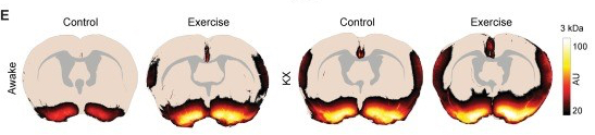
This figure shows CSF flow in awake (left) and anesthetized (right) mice. In both awake and anesthetized mice, 5 weeks of voluntary exercise increased CSF flow. Image adapted from [4]
With a new field of research burgeoning around the discovery of a glymphatic system, hopefully scientists can develop ways to increase glymphatic flow during aging as a protective measure against neurodegenerative disease. Until then, get lots of exercise, and next time you hit the snooze button on your alarm, feel free to use increased glymphatic flow as justification for sleeping in.
References:
- Iliff JJ, Wang M, Liao Y, Plogg BA, Peng W, Gundersen GA, Benveniste H, Vates GE, Deane R, Goldman SA, Nagelhus EA, Nedergaard M. A paravascular pathway facilitates CSF flow through the brain parenchyma and the clearance of interstitial solutes, including amyloid β. Sci Transl Med. 2012 Aug 15;4(147):147ra111. doi: 10.1126/scitranslmed.3003748. PubMed PMID: 22896675; PubMed Central PMCID: PMC3551275.
- Jessen NA, Munk AS, Lundgaard I, Nedergaard M. The Glymphatic System: A Beginner’s Guide. Neurochem Res. 2015 Dec;40(12):2583-99. doi:10.1007/s11064-015-1581-6. Epub 2015 May 7. Review. PubMed PMID: 25947369; PubMed Central PMCID: PMC4636982.
- Xie L, Kang H, Xu Q, Chen MJ, Liao Y, Thiyagarajan M, O’Donnell J, ChristensenDJ, Nicholson C, Iliff JJ, Takano T, Deane R, Nedergaard M. Sleep drives metabolite clearance from the adult brain. Science. 2013 Oct 18;342(6156):373-7. doi: 10.1126/science.1241224. PubMed PMID: 24136970; PubMed Central PMCID:
PMC3880190.
- von Holstein-Rathlou S, Petersen NC, Nedergaard M. Voluntary running enhances glymphatic influx in awake behaving, young mice. Neurosci Lett. 2018 Jan 1;662:253-258. doi: 10.1016/j.neulet.2017.10.035. Epub 2017 Oct 25. PubMed PMID:29079431; PubMed Central PMCID: PMC5696653.
Images:
Slime, Monster wiki- http://monster.wikia.com/wiki/Slime
Vascular system- https://www.britannica.com/science/blood-vessel
Lymphatic vessel- https://en.wikipedia.org/wiki/Lymphatic_vessel
Lymphatic System- https://en.wikipedia.org/wiki/Lymphatic_system
Rotating ventricles- https://en.wikipedia.org/wiki/Ventricular_system
Glymphatic system- https://en.wikipedia.org/wiki/File:Glymphatic_system_schematic.jpg
Glymphatic system in aging and disease: [2]
Exercise increases glymphatic system: [4]

Pingback: What’s happening in the brain with COVID-19? | NeuWrite San Diego
Pingback: Lymphedema in popular culture: The Bachelorette – Life