 June
03
June
03
Tags
Axolotls: First Ones to the Fountain of Youth
Introduction

If you look at today’s $50 Mexican peso (left), you won’t see the image of a past president or monument. Instead, you’ll see a lone salamander with outstretched arms and gills in the middle of a lake. For centuries, this small creature, known as the axolotl (ax-oh-lot-al), was well known to the people that inhabited its native environment of Lake Xochimilco in present day Mexico City, the only place in the world where it is found in the wild [1]. According to legend, the Aztec god Xólotl (a combination of the Nahuatl words for water and dog or beast) wanted to escape being sacrificed so he transformed himself into this small creature. However, nowadays, this one (often cute) animal can be found in dozens of research labs across the world, where scientists study its unique talent: regeneration of almost any body part. Brains, eyes, limbs, spinal cords, and more can all be grown back with ease in just a few days – a feat that us mammals are poorly suited for [2].
🎵I wanna be for-ever young🎵
At the tail end of the 18th century, a variety of zoologists began to take a keen interest in the axolotl [3]. One of the first things they noticed, and one you may have picked up on yourself, is that axolotls look both youthful and cute. Unlike other salamanders, axolotls appeared to (almost) always stay in their larval stage, showing exposed gills, a long tail fin, and the inability to move their eyelids, all traits designed to survive in water [4]. However, they soon discovered that this seemingly juvenile state was actually its mature form, and axolotls are built for life in the water. This “forever-young” appearance can be found in just a few other salamanders and is called neoteny.
Now, this doesn’t mean that these creatures are immortal, but rather that they simply do not undergo metamorphosis to change into a terrestrial form. This conversion from larval/aquatic to adult/terrestrial form that normally happens in other salamanders is thought to be a way for the animal to take advantage of the resources outside the water and avoid dangers (e.g. predators) found within it. In the case of axolotls, their habitat of Lake Xochimilco historically did not have any predatory fish(as will be discussed later, this is no longer the case), giving axolotls no incentive to venture out onto dry land [1].
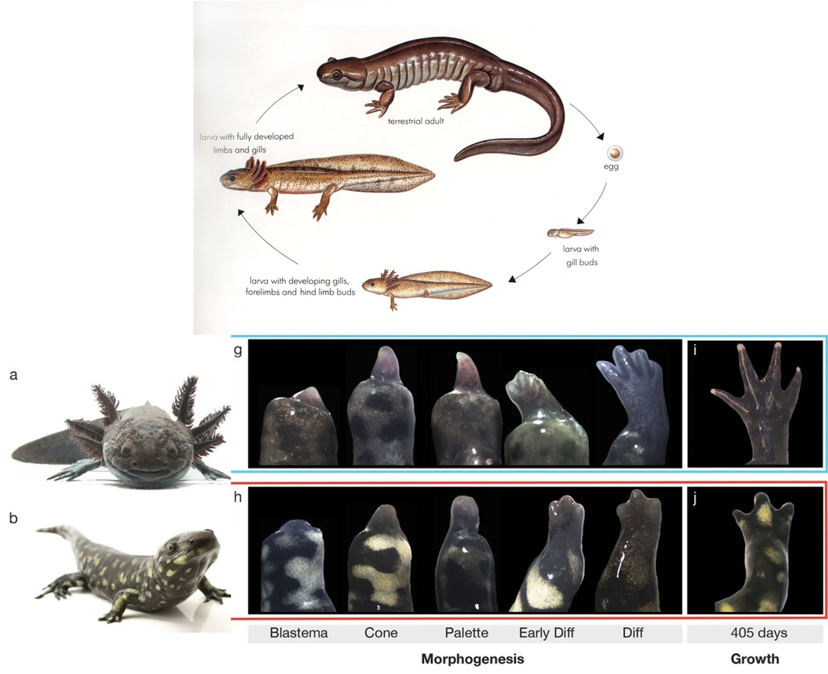
Interestingly, even though axolotls do not normally undergo metamorphosis, they still retain the ability to do so. If they become stressed and are forced to breathe air repeatedly, they can change into their adult terrestrial form. However, doing so restricts the axolotl’s ability to regenerate by about half, suggesting that neoteny is beneficial for tissue regeneration (Fig. 1) [5].
Limb regeneration
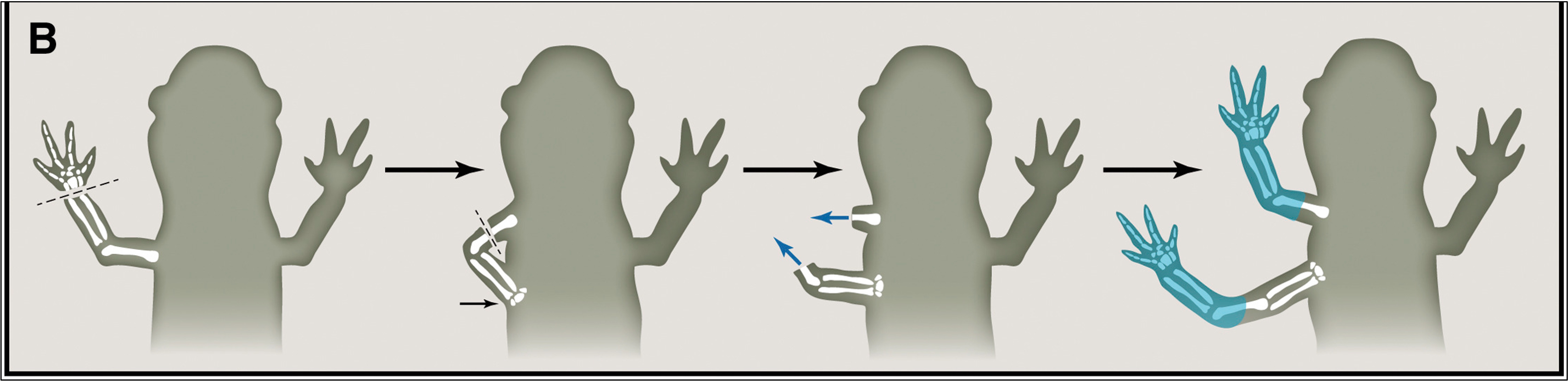
What axolotls are best known for is their ability to regenerate damaged tissue. If an axolotl limb becomes severed, it will, without fail, grow a new limb in its place. If you take a severed axolotl limb, cut off the distal end of it, and attach it to somewhere else on the body (say, to the belly), it will not only regrow a limb there, but it will regrow the exact same limb that was cut (e.g. a front right arm, or a back left leg) (Fig. 2)[6]. This small distinction may not seem like much, but it reveals a fascinating fact about the cells of an axolotl: they “know” what/where they are. Think of it like this: if I hand you bone cells from my left hand and bone cells from my right hand, they will look identical to you even under the most stringent experimental analysis. But somehow, an axolotl’s severed limb is able to make this distinction and “knows” exactly what body part to regrow. Scientists call this positional identity.
When organisms are first being formed, they use different concentrations of molecular cues to direct different groups of cells to become a specific tissue or part of a tissue (e.g. group X becomes an index finger, group Y becomes a pinkie, etc.). Researchers have found that the interplay between the secreted signaling proteins FGF and Shh on opposite ends of a developing limb play a vital role in determining the fate (e.g. index or pinkie finger to reuse the last example) of the cells that reside in those opposite ends. However, these molecular cues aren’t present in their normal watery environment, meaning that if an axolotl wants to regrow an arm, it must produce these cues itself again in adulthood.
After an amputation, a portion of the axolotl’s injured limb cells will begin to proliferate and migrate towards the injured end. Research from Dr. Elly Tanaka’s group in Vienna suggests that the very act of breaking open blood vessels during an amputation releases molecules into the injured limb that kickstart this process of cell migration and division [7]. Eventually these cells will form what is called a blastema, from which a regenerating limb will grow. Interestingly, axolotl nerves secrete several factors after being cut that are necessary for the sustained functioning of the blastema, and removing nerves prior to an amputation will prevent regeneration [6]. After making their way to the distal end, the newly migrated cells of the blastema begin secreting FGF and Shh on its anterior and posterior portions (respectively), effectively recreating the molecular cues found when the limb was first being formed. Disrupting this process can lead to improper regeneration or growth of extra limbs. It is too complicated to explain how exactly this happens, but you can read more about it in a 2016 review written by Dr. Tanaka herself, titled “The Molecular and Cellular Choreography of Appendage Regeneration [6].”
Nervous system regeneration
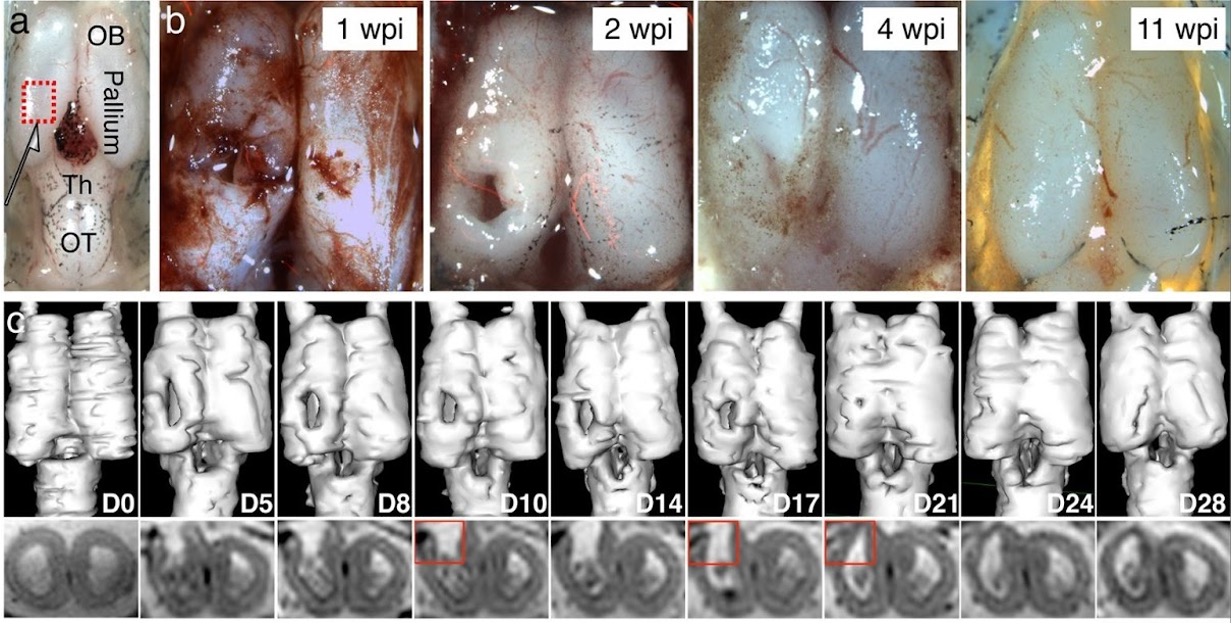
As a neuroscientist myself, I am most fascinated by the regenerative ability of the axolotl’s central nervous system. After undergoing a spinal cord injury, most mammals develop an internal scar that stops further damage from occurring and simultaneously prevents regeneration of the damaged tissue [2,8]. In axolotls, however, no scar is formed and cells all along the nervous system are programmed to quickly coordinate to repair the site of injury once it occurs. Additionally, researchers found that axolotls were able to regrow a large section of the brain that was surgically removed within a matter of weeks (Figure 3) [9]. The regenerated portions of the brain were functional (e.g. able to send signals) and included the variety of cell types you would expect to find. The only thing lacking was long-distance connections to other brain regions, suggesting that although axolotls are really good at this sort of thing, they aren’t perfect. It is well known that there is only so much brain tissue (about one hemisphere) that can be damaged before the axolotl is unable to regenerate it. How the axolotl manages to do all this and why mammals seemed to have lost this ability during the process of evolution is yet to be known.
Axolotl Endangerment
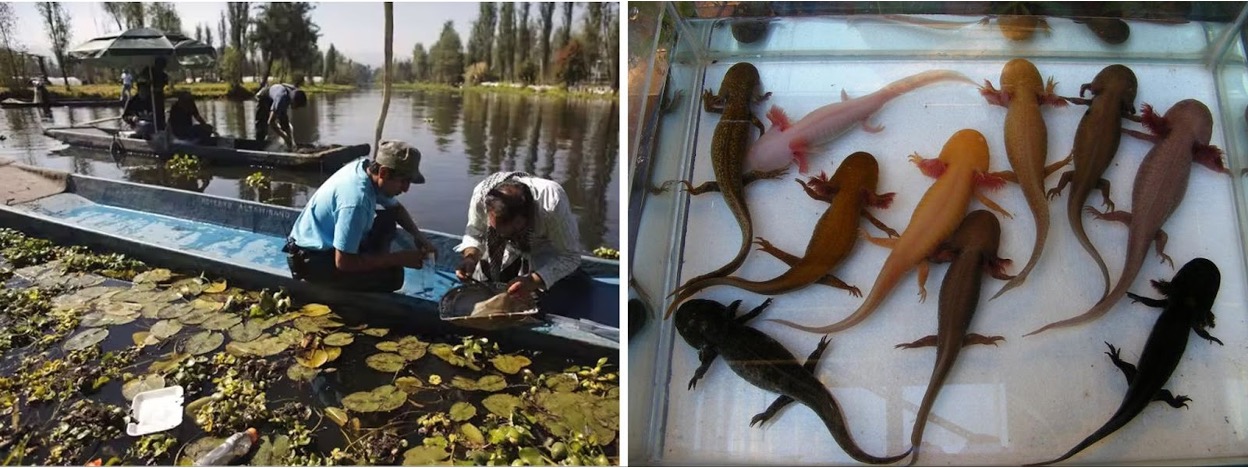
At the end of the Aztec legend of Xólotol, Quetzalcoatl, Xotol’s twin brother, finds him in his new aquatic form and gives him a stern warning, “Since you failed to sacrifice yourself to give life to another, you will spend your whole life here. But the day that your life, the water, no longer works, you will disappear from the face of the Earth [1].” Sadly, it seems that this warning is coming to fruition. In the days since the Aztec empire fell, Lake Xochimilco has been reduced to a small fraction of its size into what are essentially only canals. To make matters worse, the burgeoning metropolis of Mexico City began using more and more water from the lake’s natural sources, leading to its replenishment to come from artificial and polluted sources. Furthermore, carp and tilapia were introduced in the 1970s to stimulate an aquaculture economy, but subsequently led to axolotl eggs and young axolotls being eaten before they could reproduce. The most grim predictions state that axolotls will be fully extinct in the wild by 2025 [1]. Restoration projects are underway by conservationists and academics, often using the introduction of axolotls bred in research labs to new sanctuary lakes that have cleaner sources of water and lack predatory fish. Even though this will not be the magnificent return of the axolotl to its home habitat, it will allow this wonderful creature to continue living in the wild outside of academic labs.
References
- Hernandez Mares, P., 2017. Ajolote: el hermano de Quetzalcóatl que pelea por sobrevivir. [online] Cientificodigital.mx. Available at: <https://cientificodigital.mx/articulos/naturaleza/44/ajolote-el-hermano-de-quetzalcoatl-que-pelea-por-sobrevivir> [Accessed 11 May 2022].
- Chernoff, E. A. G., Stocum, D. L., Nye, H. L. D. & Cameron, J. A. Urodele spinal cord regeneration and related processes. Dev. Dyn. 226, 295–307 (2003).
- Reiß, C., Olsson, L. & Hoßfeld, U. The history of the oldest self-sustaining laboratory animal: 150 years of axolotl research. J. Exp. Zool. Part B Mol. Dev. Evol. 324, 393–404 (2015).
- De Groef, B., Grommen, S. V. H. & Darras, V. M. Forever young: Endocrinology of paedomorphosis in the Mexican axolotl (Ambystoma mexicanum). Gen. Comp. Endocrinol. 266, 194–201 (2018).
- Monaghan, J. R. et al. Experimentally induced metamorphosis in axolotls reduces regenerative rate and fidelity. Regeneration 1, 2 (2014).
- Tanaka, E. M. The Molecular and Cellular Choreography of Appendage Regeneration. Cell 165, 1598–1608 (2016).
- Currie, J. D. et al. Live Imaging of Axolotl Digit Regeneration Reveals Spatiotemporal Choreography of Diverse Connective Tissue Progenitor Pools. Dev. Cell 39, 411 (2016).
- Costa, E. C., Otsuki, L., Albors, A. R., Tanaka, E. M. & Chara, O. Spatiotemporal control of cell cycle acceleration during axolotl spinal cord regeneration. Elife 10, (2021).
- Amamoto, R. et al. Adult axolotls can regenerate original neuronal diversity in response to brain injury. Elife 5, (2016).
- https://www.independent.co.uk/climate-change/news/axolotl-found-in-mexico-city-lake-after-scientists-feared-it-only-survived-in-captivity-9148775.html

You must be logged in to post a comment.