 October
04
October
04
Tags
The ethics of human brain surrogacy
“Creepy ‘brain in a bucket’ study spurs medical, ethical debates” … “Yale experiment to reanimate dead brains promises ‘living hell’ for humans” … “Scientists have managed to reanimate disembodied pigs’ brains – but for a human mind, it could be a living hell” … These are just a few of the sensational headlines that came out earlier this year, when a group of neuroscientists took about 200 brains from recently decapitated pigs and kept them “alive” with a system of pumps, heaters, and artificial blood. The lead scientist of this group, Dr. Nenad Sestan, described the work earlier this year at an ethics workshop at the National Institute of Health (NIH), and has declined to comment any further on the study until their scientific report is published. Nonetheless, the few details we have about this project have spurred an outpour of excitement and concern among scientists and non-scientists alike.
Brain in a bucket
So what do we know about this “brain in a bucket” study, officially termed BrainEx? The research team started the project with the goal of creating a comprehensive map of all the connections between cells in the human brain. Keeping an entire, intact brain alive outside of the body would be a huge aid in this goal. According to Sestan, the BrainEx system was able to restore circulation to blood vessels throughout the brains and keep individual neurons active and capable of “normal” activity for up to 36 hours. This is the first time a large mammalian brain has been kept active, completely separated from the rest of the body (another successful attempt occurred in 1993, when a research team kept a guinea pig brain alive in a similar fashion for 8 hours [1]). This is a huge breakthrough for brain researchers; if this system can be replicated with other animals, we may be able to study the brain as a complete, functioning organ in ways that we haven’t been able to before.
However, since the news of this study broke, people everywhere have begun voicing concerns over the ethical implications of such a system. Headlines, such as those at the beginning of this article, reveal fears that the animals which the brains belonged to may be conscious as their brain is kept active. However, Sestan and his team recorded electrical activity from the pig brains, and found broad patterns similar to those seen in a comatose brain. Nonetheless, Sestan commented that he believes it could be possible to restore consciousness and keep ex-vivo brains alive indefinitely, but he and his team did not want to step into such uncharted territory.
Indeed, Sestan has clearly shown his concern over the ethical implications of expanding such a technique to human subjects. Although we are far from being able to maintain a human brain outside of the body, our tools for studying the brain are becoming more sophisticated. About a month after the ethics workshop at the NIH, Sestan along with 16 other scientists published an essay in the journal Nature titled “The ethics of experimenting with human brain tissue” [2]. In this essay, the researchers discuss three different surrogates for human brains which are becoming commonly used in laboratories today: ex-vivo brain tissue, organoids, and chimaeras.
Options for human brain surrogacy

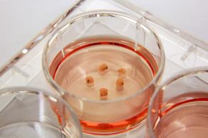
ex-vivo human brain tissue [2]
brain organoids [2]
Finally, chimaeras are organisms that contain genetic material from two different animals (appropriately named after a mythological fire-breathing monster with a lion’s head, a goat’s body, and a snake’s tail!). Although this sounds like something you would read about in a sci-fi novel, chimaera research is actively being pursued. This technique can be used to examine how cells from one animal can become integrated and function in another, and has exciting biomedical implications. For example, someday we may be able to grow organs from patient-derived stem cells in animals and transplant them into patients in need.
The authors argue that, with the current state of research, it is highly unlikely that these three types of human brain surrogates could experience human-like consciousness or awareness of any kind. However, they argue that as these brain models become more advanced, clear ethical guidelines need to be established in order to address issues such as consent, stewardship, and ownership of brain tissue.
“As brain surrogates become larger and more sophisticated, the possibility of them having capabilities akin to human sentience might become less remote. Such capacities could include being able to feel (to some degree) pleasure, pain or distress; being able to store and retrieve memories; or perhaps even having some perception of agency or awareness of self” Farahany et al. 2018 [2]
Human-mice chimaeras
Recently, a team of scientists at the Salk Institute accomplished one such advancement by combining two of these brain surrogacy methods: organoids and chimaeras [3]. A major limitation with organoids is that the bigger they are, the more difficult it is to keep them “alive”, as without blood circulation nutrients can’t make their way to the center of the tissue. The Gage lab sought to solve this problem by transplanting human brain organoids into the mouse brain. Amazingly, within a few days, the organoids showed integrated vascularization (read: blood flowing through the vessels).
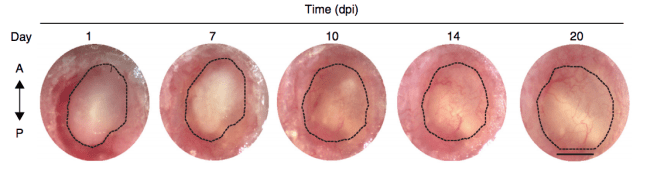
“Serial microscopic tracking of grafts showing dynamics of blood essel perfusion by the recipient vascular system” [3]
What we can do vs. what we ought to do
Of course, as promising as this line of research is, methods like these raise several ethical concerns. Particularly in the case of inserting human neural tissue into the brain of another animal, one major (and perhaps the most alarming) concern is the possibility of human-like consciousness in the animal being studied. While this is unlikely given current technology, research has already shown that similar techniques are capable of enhancing the cognitive capabilities of an animal. A study by Xiaoning Han and colleagues (2014) demonstrated that inserting a type of human brain cell, called glia, into the forebrains of mice helped these mice learn more quickly across several behavioral tasks [4]. (For a greater discussion of the ethical issues of human-animal chimaera research, see this article by Göran Hermerén.)
Thus, as we move forward with more sophisticated models of human brain surrogacy, it will be important to continue checking in with ourselves and our communities to ensure that ethical values are being considered and respected. It will be critical for the scientists behind these types of studies to communicate clearly and effectively with the public to ensure that any fears that may exist are not based on ignorance. Just as well, the general public should keep the scientific community in check to ensure that the pushing of ethical boundaries is predicated on a desire to benefit people, rather than a desire for knowledge regardless of the risks involved.
References
- Muhlethaler M, de Curtis M, Walton K, and Llinas R (1993). The isolated and perfused brain of the Guinea-pig in vitro. European Journal of Neuroscience 5, 915-926.
- Farahany NA, Greely HT, Hyman S, Koch C, Grady C, Pasca SP, Sestan N et al. (2018). The ethics of experimenting with human brain tissue. Nature Comment 556, 429-432.
- Mansour AA, Goncalves JT, Bloyd CW, Li H, Fernandes S, Quang D, Johnston S et al. (2018). An in vivo model of functional and vascularized human brain organoids. Nature Biotechnology 36(5), 432-441.
- Han, X., Chen, M., Wang, F., Windrem, M., Wang, S., Shanz, S., Xu, Q., Oberheim, N. A., Bekar, L., Betstadt, S. et al. (2013). Forebrain engraftment by human glial progenitor cells enhances synaptic plasticity and learning in adult mice. Cell Stem Cell 12, 342-353.

Pingback: Life After Death(?): From Strokes to Sci-Fi | NeuWrite San Diego