 February
20
February
20
Trying to Forget
It is difficult to overstate the importance of learning. Many consider lifelong learning to be one of their primary goals in life, education is one of the primary roles of government, and machine learning — an algorithmic approximation of learning applied to computers — is a hundred-million dollar business. It quite literally underlies everything we know. As a result, most neuroscience students can recite the basic details of learning in their sleep. Frequently used connections (also known as synapses) between neurons active in response to an input will be strengthened, making activation of the network even easier the next time around. This idea is called Hebb’s rule or Hebbian learning, and most simply sum it up as: “Neurons that fire together wire together.” There’s a lot more to it, of course, but it’s a bit outside the scope of this article: this is not actually an article about learning.
Instead, I want to talk about its more enigmatic opponent, forgetting. Despite all the attention given to how each and every aspect of learning works, far less has been given to how we forget. Maybe it’s because we see forgetting as an annoyance to be corrected, or maybe it’s because millions of dollars aren’t dependent on machine forgetting. Just on a philosophical level, though, forgetting is far more important than most people think: without the ability to forget, our brains cannot choose which information is actually important.
On a biological level, forgetting is even more consequential. To learn new information, in most cases you must eventually forget at least some of the old info at some point, independent of its importance. The wiring between two cells after learning occurs is only strong relative to weaker connections. If you cannot reduce the strength of certain connections, the network soon becomes saturated at maximum strength. When this happens, we actually lose the ability to store new memories entirely [1].
How do we forget?
At first, most scientists thought that we did not actually remove the information we forget. Instead, errors happen over time, and you slowly become unable to retrieve the memory from storage. Under this view, reminders can easily reinstate the network, as you do not have to re-learn everything, just the few parts lost. However, this view violates nearly everything we know about biology. When a part of a cell is no longer necessary, like a used protein, the cell does not just allow it to hang around until it falls apart. Instead, it swiftly deals with the problem using components specifically designed for the task. Such active maintenance is often extremely costly, but nearly always required for optimal function. If such processes were not active in the brain, it would be very odd indeed.
In response, a cadre of neuroscientists introduced the concept of “active forgetting”, where neurons actively weaken certain connections that are less active than others [2]. Through a variety of cleverly done experiments in a series of high-profile studies, they were able to show not only that this active forgetting occurs, but it uses a protein involved in changing synaptic strength , known as Rac1. For decades, Rac1 has been thought to be crucial for learning, changing the structure of synapses in response to activity. However, by altering the protein, they were able to show that it does become activated by similar processes to learning, and it does change the structure of the synapse. Instead, the direction was reversed: in their studies, Rac1 instead broke down the synapse instead of strengthening it, causing the connection to decay if too weak. According to the theory, this protein can induce decay at specific synapses, causing certain memories to be forgotten.
However, a number of issues exist with this model, too. First of all, forgetting usually does not involve re-activating the same neurons involved in the initial memory, which would be required under this model. Instead, this process needs to occur independent of stimulation, and such a vehicle for forgetfulness has not yet been found. Some early evidence points to a potential role for sleep, but far more research is required before we can confidently make that conclusion. In the interim, a second model has begun to form, centered on a set of cells relatively obscure until recently: microglia.
Microglia?
Microglia are small (“micro”) cells in the nervous system that do not themselves transmit or store information (these cells are known as “glia”), but instead defend the brain from infection. For the most part, they are the immune system in the brain, and can be thought of as similar to white blood cells. For a while, they were seen as a boring topic to study. If you were to ask a neuroscientist 20 years ago, they would likely say that microglia are just white blood cells in the brain. They just destroy foreign invaders, right?
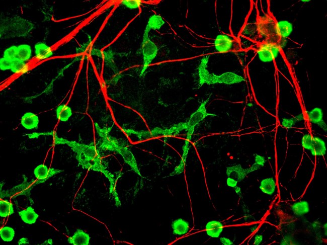
Microglia (green) and neurons (red) in a dish. (Wikimedia Commons)
Wrong. In the past ten years, our ideas about the function of microglia have changed dramatically. First, two papers published around 2010 made a fundamental observation not previously on anyone’s radar [3,4]. By taking videos of microglia moving within the brain, they found that microglia did not just search the brain for invaders, but also for neural activity. Microglia would frequently contact synapses, and would change how often the contacts occurred if the neurons were active. Further, microglia did not stop at touching the synapse, but would frequently destroy it afterwards. In other words, microglia examine how active the surrounding synapses are, and in many cases destroy them afterwards, though the scientists did not know why. However, this question was quickly answered in a follow-up study two years later, where a group led by Dr. Beth Stevens at Harvard found that activity and synaptic destruction were indeed linked: increases in neural activity protected against synaptic destruction by microglia, and reductions made synapses more susceptible to pruning [5].
Though both synaptic pruning and its regulation by activity both point to microglia being involved in memory, no one had yet actually proved it until 2016. That year, Beth Stevens’ lab again published two papers showing that two diseases linked to memory loss, West Nile Virus and Alzheimer’s disease, both involved microglial activation via the complement cascade, a system that both recruits immune cells to targets and destroys them directly [6,7]. Before these papers were released, most thought that memory loss was an intrinsic property of the disease. Instead, memory loss in these diseases may instead possibly be a result of (or at the very least made worse by) microglia trying to fight off the disease, destroying synapses in the process. These two studies both upended how we think of these respective diseases and linked microglia to memory decline in disease by a very specific mechanism, but the story was still incomplete.
A few weeks ago, that first chapter of that story finally reached its conclusion [8]. Dr. Yan Gu, a professor in Hangzhou, China, was able use contextual fear conditioning, a standard technique to teach mice to associate certain surroundings with pain. Using a drug known as PLX3397, an experimental treatment to selectively kill microglia in the brain, he was able to examine microglia’s contribution to memory. By treating a subset of the mice with this drug for 5 weeks, the mice could forget their fearful experience, with or without microglia. Astonishingly, the PLX-treated mice that had their microglia killed didn’t forget! Further, when they caused neuron storing the memory to express parts of the complement cascade, most of their synapses were destroyed in the presence of microglia. Finally, when synaptic activity was suppressed while in the presence microglia, the memory-storing synapses were all destroyed, and the mice forgot, which did not happen when microglia were removed. In other words, microglia control forgetting by monitoring neural activity and selectively eliminating synapses using the complement cascade.
Though these studies are provocative, a number of questions remain unanswered. First, how do microglia actually detect neural activity? All of these results are built on observations of microglia’s proximity to active synapses and pruning that happens afterwards. These observations do not include the mechanism by which it happens, or what microglia actually use to detect activity. Further, no one knows how active a synapse must be to survive, which could have major ramifications for how the process works overall. Finally, do neurons send signals to microglia themselves to highlight synapses to destroy, or do microglia do it all on their own? Either possibility could open up the door to many therapies for a wide range of diseases with cognitive effects, such as lupus or AIDS.
Regardless, these studies radically redefine not only how we think of forgetting, but of how we think of the brain as a whole. Instead of the mind being a creation of our neurons alone, thought may be a more cooperative process than expected. Neurons may not be able to independently forget information, but instead could require cells like microglia to identify unnecessary synapses and remove them. Though these results may be limited to forgetting for now, it opens the door for re-evaluation of numerous mental processes through a whole new lens.
Though these above findings may change our understanding of microglia, which of these theories of forgetting are correct? Though all three (memory decay, active forgetting, and microglial forgetting) work in dramatically divergent ways, each is different enough where one does not actually prevent the others from being true either. Sure, it could be one or it could be the other, but it could also be both, or it could also be neither. This uncertainty speaks to the relative neglect towards research on forgetting, and the field needs to do far more work before we can decide conclusively. No matter what future scientists may find, the results will surely be unforgettable.
References:
- Moser, E.I., Krobert, K.I., Moser, M.B., Morris, R.M. (1998). Impaired Spatial Learning after Saturation of Long-Term Potentiation. Science. 281:2038-2042.
- Davis, R.L., Zhong, Y. (2018). The Biology of Forgetting – A Perspective. Neuron. 95:490-503.
- Tremblay, E.M., Lowery, R.L., Majewska, A.K. (2010). Microglial Interactions with Synapses Are Modulated by Visual Experience. PLoS Biol. 8(11): e1000527.
- Wake, H., Moorhouse, A.J., Jinno, S., Kohsaka, S., Nabekura, J. (2009). Resting Microglia Directly Monitor the Functional State of Synapses In Vivo and Determine the Fate of Ischemic Terminals. J Neurosci. 29:3974-3980.
- Schafer, D.P., Lehrman, E.K., Kautzman, A.G., Koyama, R., Mardinly, A.R., et al. (2013). Microglia Sculpt Postnatal Neural Circuits in an Activity and Complement-Dependent Manner. Neuron. 74:691-705.
- Vasek, M.J., Garber, C., Dorsey, D., Durrant, D.M., Bollman, B., et al. (2016). A complement–microglial axis drives synapse loss during virus-induced memory impairment. Nature. 534:538-543.
- Hong, S., Beja-Glasser, V.F., Nfonoyim, B.M., Frounin, A., Li, S., et al. (2016). Complement and microglia mediate early synapse loss in Alzheimer mouse models. Science. 352:712-716.
- Wang, C., Yue, H., Hu, Z., Shen, Y., Ma, J., et al. (2020). Microglia mediate forgetting via complement-dependent synaptic elimination. Science. 367:688-694.

Pingback: Microglia as Architects and Designers for Your New Brain | NeuWrite San Diego