 December
04
December
04
Controlling the brain with lasers!
Two male mice are hanging out in a cage, both with strange looking cables coming out of their head, and both minding their own business, when all of a sudden, a researcher flips a switch, sending a green light down the cable and into one of the mice’s brain. The mouse immediately proceeds to attack the second, with seemingly no provocation, but immediately stops when the researcher turns the light off. Some time passes, the mice are fighting, as mice sometimes do. The researcher flips a different switch, this time sending a red light into the brain of the other mouse, which immediately stops fighting. The researcher turns off the light and the mouse resumes fighting. This has been a demonstration of a trendy technique in neuroscience: optogenetics, controlling individual brain cells with finely-tuned wavelengths of light. These mice both have fiber optics planted deep in their brains, in a part of ventromedial hypothalamus. The first mouse has a specific channel that excites the neurons in response to green light, whereas the other has red light-responsive channels that silence the neuron when activated. This study effectively showed that by taking control of neuronal firing in this region, they were able to control aggression in these mice (1).
How does it work?
So how does this work, anyways? Controlling the brain with lasers sounds impossibly science fiction-y, but the technology behind it is actually fairly straightforward. In order to understand how optogenetics is controlling a neuron, we must first have a basic understanding of how neurons communicate. Essentially, neurons relay signals from their dendrites (which contact other neurons) to the cell body, which sum up these signals and fire an action potential. These action potentials are a brief change in the electric potential of the cellular membrane, a change which travels towards the cell body and is caused by the opening and closing of ion channels. From the cell body, the action potential moves down the axon, which will send signals to other neurons. Optogenetics works by making the neuron express an ion channel from algae, channelrhodopsin, which opens in response to light. This opsin is then incorporated in the neuron’s membrane, and a specific wavelength of light will prompt the opsin to open its ion channel, causing a depolarization of the membrane, effectively causing an action potential. The light comes from either a laser via an optical fiber that runs into the brain, or a surgically implanted LED.
Since the technology’s inception, a whole slew of other opsins and similar molecules have been developed. By using different opsins, we can either depolarize the membrane, causing the neuron to fire, or hyperpolarize it, effectively preventing the neuron from sending any action potentials. These opsins are stimulated by different wavelengths of light, so they can be put in the same neuron, allowing us to effectively turn the neuron on and off at will. Other ion channels have been developed that are able to open and close much faster, mimicking the higher firing rate of some neurons, and a new wave of proteins modify protein-protein interactions in response to light, allowing us to control different cell types in a whole new way. A new method, dubbed InSynC, even controls synaptic vesicle release, which is how the axon of one neuron communicates to another neuron across the synapse (2).

They can control your synaptic vesicle release with lasers!
In order to express a light-sensitive protein in neurons, a genetic construct containing the opsin and a protein that fluorescently tags the cell (in order to identify where it is expressed) is placed in a virus that is then injected into the region of interest in the brain. Importantly, the type of virus that is used is from the same family as HIV and, like HIV, is able to incorporate itself into the genome of the cell, hijacking the cell’s own mechanisms in order to express the opsin and reporter. Fortunately, the virus has been modified in such a way that it is unable to replicate and take over the cell.
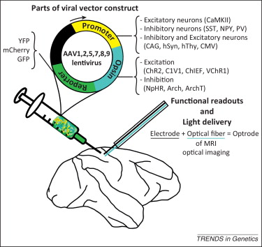
In order to optogenetically activate a region, a viral construct must first be injected, then an optogenetic probe lowered into the region of interest. From Gerits and Vanduffel 2013
To control the cells in which the opsin is expressed, the regulatory elements (promoter) of a cell-type specific gene will be put in control of the opsin’s expression. Often, the expression of the opsin will be dependent on the expression of a particular gene inserted into the genome of an animal that marks a specific cell type (as in the cre-lox system), a more effective method, albeit dependent on the availability of transgenetically manipulated animals. In both systems, the opsin will only be expressed where that gene is expressed, enabling specific types of neurons to be manipulated. This methodology is key to the success of optogenetics: by controlling subsets of neurons in a region, we can parse apart the role of different neuronal networks and systems. Regions of the brain consist of many different cell types close together that receive different inputs and have different outputs, sometimes doing completely opposite things, such as the case of inhibitory versus excitatory neurons.
Applications of optogenetics: the cool and the practical
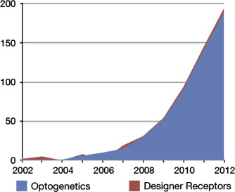
The growth of optogenetics in published research studies. From Aston-Jones and Deisseroth, 2013
Optogenetics has been trendy in neuroscience since its first successful demonstration by Karl Deisseroth in 2005 “Ever since Karl Deisseroth and his post-doc Ed Boyden first successfully controlled mammalian neurons with lasers in 2005, optogenetics has become an incredibly trendy neuroscience tool, as shown in this first figure (for an interesting history of optogenetics’ development, which I’m not going to go into here, read this piece by Boyden). Now, its use is practically mandatory if you want to prove that a set of neurons has a particular function in the brain. This isn’t surprising; optogenetics allows us to control how specific neurons fire on precise time scales, allowing us unprecedented control of the brain. Plus, lasers are sexy. So far, optogenetics has been applied to everything from neurons in a dish, fly brains, and roundworms, to mice, primates, and practically a whole zoo of other systems–though rodent work remains most prevalent. So what have people done with it?
- In the roundworm, each of its 302 neurons can be activated individually, or in particular subsets. Several studies have shown that activation of a particular neuron results in forward acceleration, whereas stimulating a different neuron results in the reverse (4) (as to what activating both at the same time will do, I don’t know: maybe the worm will explode?).
- A study in fruit flies found that activating a set of 12 neurons evoked what amounts to a fear memory; optogenetically activating these neurons resulted in the flies avoiding where they happened to be when the laser turned on (5).
- Recent work implanted a false fear memory in mice by activating a subset of neurons in the hippocampus, an area strongly associated with memory. Here, they activated neurons that were shown to be involved a traditional fear conditioning paradigm (a foot shock) in a context outside of the paradigm and found that this unconditioned but light-stimulated mouse responded to the laser-associated context the same way as the fear-associated one (6)
- Optogenetics is also capable of both causing and relieving Obsessive-Compulsive Disorder-like symptoms in mice. Light-mediated activation of a circuit shown to be dysregulated in human OCD patients caused OCD-like symptoms in mice (which involves persistent grooming, among other things) (7). A different lab (in the same issue of Science) showed that optogenetically stimulating a different, yet related, circuit in the mouse model of OCD reduced OCD-like behaviors to baseline levels (8).
These are only a few of the hundreds of contexts where optogenetics has been used to a myriad of effects: causing, stopping, or altering countless behaviors.
More than just neurons
While this technology was developed in the brain for neurons, it’s not confined to neurons or the brain. Sergey Kasparov’s lab found that by optogenetically stimulating astrocytes–a different, highly abundant cell type found in the brain and spinal cord–in the brainstem they were able to control breathing (9). Additionally, using the same principles as in neurons, optogenetics has been used to control heartbeat (10), with pulses of light essentially pacing the firing of heart cells, telling them when to contract. This works because most muscle cells communicate through electric impulses traveling along their membrane, just as neurons do. Theoretically, this means that we can control movement with light.
Limits
Like any technology, optogenetics has its limits. First and foremost, we are limited by the neurons we can mark. Not all cell types have known genetic markers, and if we can’t mark the cell type genetically, it is much harder (and sometimes impossible) to insert an opsin into those cells alone. We also need to know enough about different circuits and cell types in the brain in order to target them, which is no small feat. Beyond that, only a handful of animals have the established, pliable genetics required for making genetically manipulated animals that make it easy to successfully interrogate a given cell type. While the development of new technologies such as the excellently named TALENS and its less welled-named but more likely to succeed cousin CRISPR will make it easier to manipulate genomes across the board, the fact remains that it is much easier to excite your cells of interest in mice or roundworms than it is in leeches or monkeys. There are also limitations in being able to effectively mimic neuron firing. Different neuronal cell types, of which there are many, fire in widely different patterns, with different electrical signatures, both of which vary in response to the stimuli received. How you are controlling the input is then quite important, and often overlooked, as these parameters may be unknown. It’s also hard to gauge the expression of the inserted opsin. The efficacy of viral infection varies significantly, making it easy to over-express the opsin and overexcite the neuron, causing ion depletion and effectively silencing the neuron. Conversely, too little viral expression will lead to no effect at all.
The Future

Ohmygodmarmosetyouarecute
While a bunch of cool work will undoubtedly continue to happen in other model systems the application of optogenetics to the monkey brain is the next frontier for this technology. Less than a half dozen studies have induced behavioral responses from optogenetic stimulation in primates, due to the expense and difficulty in targeting subsets of neurons in the monkey. Genetic manipulations of the sort that are standard in mice, which enable the specificity of opsin integration to subsets of cells, simply don’t exist in the primate, though there hasbeen a recent push to develop genetically manipulated (transgenic) primates, most promisingly with marmosets. Marmosets are more amenable to being raised in lab environments and have a relatively quick generation time, yet are still quite genetically similar to humans, with important similarities in brain architecture and genetics that other non-primate model organisms lack (11) By offering fine control of neuronal firing, optogenetics holds much promise in interrogating the brains of our close animal relatives.
Of course the holy grail of optogenetics, or nightmare, depending on your perspective, is its use in humans. Countless therapies could be imagined and possibly developed using optogenetics, though we certainly are limited by the fact that transgenic humans are out of the question. New ways of targeting neuronal subsets will have to be developed that don’t rely on genetic manipulation, such as some sort of “gene therapy”, but many of the viral vectors already used in optogenetics are already FDA-approved.13 There is even some precedence for modulating human neurons in an even cruder way. Deep Brain Stimulation (DBS), which uses an electrode to stimulate a specific region of the brain, is an approved, albeit radical, treatment for epilepsy and Parkinson’s, and is in clinical trials to treat major depression. This could be replaced by optogenetics, which, even if not targeted to specific neurons, is probably a safer, more effective alternative to electrodes in one’s brain. Even if we are able to get over safety issues and technical hurdles, we will still be limited by our knowledge of the neuronal underpinnings of mental conditions. It’s all well and good to be able to modulate the firing of specific neurons, but we first need to know which neurons are firing abnormally, which is something a long way off in humans for most mental disorders. Hopefully, in using optogenetics with organisms from roundworm to the marmoset, we can elucidate exactly what’s going on in that fleshy bit between our ears.
References
1) Lin D, Boyle MP, Dollar P, Lee H, Lein ES, Perona P, Anderson DJ. Functional identification of an aggression locus in the mouse hypothalamus. Nature. 2011 Feb 10;470(7333):221-6. doi: 10.1038/nature09736.
2) Lin JY, Sann SB, Zhou K, Nabavi S, Proulx CD, Malinow R, Jin Y, Tsien RY. Optogenetic inhibition of synaptic release with chromophore-assisted light inactivation (CALI). Neuron. 2013 Jul 24;79(2):241-53. doi: 10.1016/j.neuron.2013.05.022.
3) Aston-Jones G, Deisseroth K. Recent advances in optogenetics and pharmacogenetics. Brain Res. 2013 May 20;1511:1-5. doi: 10.1016/j.brainres.2013.01.026.
4) Husson SJ, Gottschalk A, Leifer AM. Optogenetic manipulation of neural activity in C. elegans: from synapse to circuits and behaviour. Biol Cell. 2013 Jun;105(6):235-50. doi: 10.1111/boc.201200069.
5) Claridge-Chang A, Roorda RD, Vrontou E, Sjulson L, Li H, Hirsh J, Miesenböck G. Writing memories with light-addressable reinforcement circuitry. Cell. 2009 Oct 16;139(2):405-15. doi: 10.1016/j.cell.2009.08.034.
6) Ramirez S, Liu X, Lin PA, Suh J, Pignatelli M, Redondo RL, Ryan TJ, Tonegawa S. Creating a false memory in the hippocampus. Science. 2013 Jul 26;341(6144):387-91. doi: 10.1126/science.1239073.
7) Ahmari SE, Spellman T, Douglass NL, Kheirbek MA, Simpson HB, Deisseroth K, Gordon JA, Hen R Repeated cortico-striatal stimulation generates persistent OCD-like behavior. Science. 2013 Jun 7;340(6137):1234-9. doi: 10.1126/science.1234733.
8) Burguière E, Monteiro P, Feng G, Graybiel AM Science. Optogenetic stimulation of lateral orbitofronto-striatal pathway suppresses compulsive behaviors. 2013 Jun 7;340(6137):1243-6. doi: 10.1126/science.1232380.
9) Gourine AV, Kasymov V, Marina N, Tang F, Figueiredo MF, Lane S, Teschemacher AG, Spyer KM, Deisseroth K, Kasparov S. Astrocytes control breathing through pH-dependent release of ATP. Science. 2010 Jul 30;329(5991):571-5. doi: 10.1126/science.1190721.
10) Bruegmann T, Malan D, Hesse M, Beiert T, Fuegemann CJ, Fleischmann BK, Sasse P. Optogenetic control of heart muscle in vitro and in vivo. Nat Methods. 2010 Nov;7(11):897-900. doi: 10.1038/nmeth.1512.
11) J Mashiko H, Yoshida AC, Kikuchi SS, Niimi K, Takahashi E, Aruga J, Okano H, Shimogori T. Comparative anatomy of marmoset and mouse cortex from genomic expression. J Neurosci. 2012 Apr 11;32(15):5039-53. doi: 10.1523/JNEUROSCI.4788-11.2012.
12) Nayerossadat N, Maedeh T, Ali PA. Viral and nonviral delivery systems for gene delivery. Adv Biomed Res. 2012;1:27. doi: 10.4103/2277-9175.98152.
13) Gerits A, Vanduffel W. Optogenetics in primates: a shining future? Trends Genet. 2013 Jul;29(7):403-11. doi: 10.1016/j.tig.2013.03.004.

Here’s a history of optogenetics from Ed Boyden himself 🙂 http://syntheticneurobiology.org/publications/publicationdetail/136/25
Pingback: A Toast to Optogenetics | NeuWrite San Diego
Pingback: Dawn of the DREADD | NeuWrite San Diego
Pingback: How Neuroscience Tools Can Help Patients Regain Their Vision | NeuWrite San Diego